ATLAS OF RADIOGRAPHIC INTERPRETATION

in small animals
IMAGES OF NORMAL RADIOGRAPHIC ANATOMY
ISABEL GARCÍA REAL - Diagnostic Imaging Service, Veterinary Teaching Hospital, Complutense University of Madrid - PIMCD 256 (2011-2012) - UCM

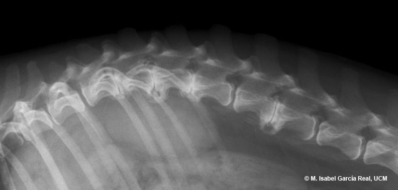
Radiological report

Taking into account the patient’s clinical picture, these findings suggest degenerative disc disease, with possible protrusion/extrusion of one or several discs at different degrees. The use of magnetic resonance imaging, computed tomography or myelography (in order of preference) is required to confirm the diagnosis and determine the exact points and the severity of the possible spinal cord compression.

Previous
Centro Empresarial El Trovador, planta 8, oficina I - Plaza Antonio Beltrán Martínez, 1 • 50002 Zaragoza - España • Tel.: 976 461 480 • Fax: 976 423 000 • www.grupoasis.com