ATLAS OF RADIOGRAPHIC INTERPRETATION

in small animals
IMAGES OF NORMAL RADIOGRAPHIC ANATOMY
ISABEL GARCÍA REAL - Diagnostic Imaging Service, Veterinary Teaching Hospital, Complutense University of Madrid - PIMCD 256 (2011-2012) - UCM

Radiological report

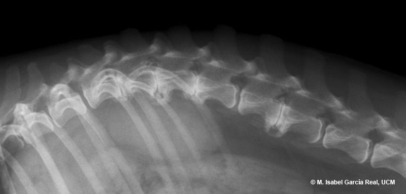
Radiograph of the thoracolumbar spine in lateral projection.


The following radiological findings are observed:


- Narrowing of the T12-T13, T13-L1, L1-L2 and L3-L4 intervertebral disc spaces. These vertebrae are also affected by spondylosis deformans at different degrees.


- Mineral density shadows located immediately dorsal to the T13-L1 (as a double arch), L3-L4 (two oval shadows) and L4-L5 (one long oval shadow) intervertebral disc spaces.


Centro Empresarial El Trovador, planta 8, oficina I - Plaza Antonio Beltrán Martínez, 1 • 50002 Zaragoza - España • Tel.: 976 461 480 • Fax: 976 423 000 • www.grupoasis.com