ATLAS OF RADIOGRAPHIC INTERPRETATION

in small animals
IMAGES OF NORMAL RADIOGRAPHIC ANATOMY
ISABEL GARCÍA REAL - Diagnostic Imaging Service, Veterinary Teaching Hospital, Complutense University of Madrid - PIMCD 256 (2011-2012) - UCM

Radiological report

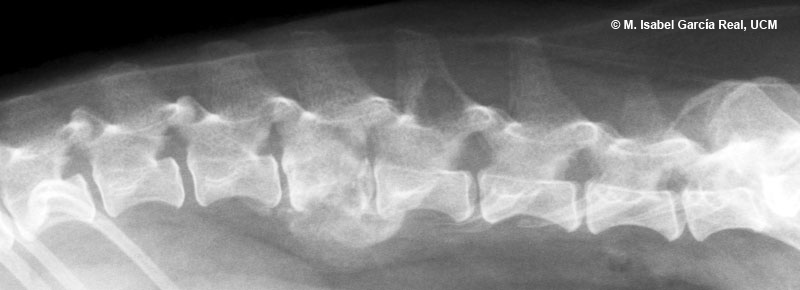
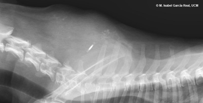
Radiographs of the cervicothoracic and lumbar spine in lateral projections.


A superficial soft-tissue mass is observed in the cervicothoracic region. It is located dorsal to the spine and extends from the level of C4 to T3. The mass has small areas of calcification and the identification microchip is superimposed on its central area. Small-sized osteolytic lesions are observed in the spinous processes of C4, C5, C6 and T6.


Centro Empresarial El Trovador, planta 8, oficina I - Plaza Antonio Beltrán Martínez, 1 • 50002 Zaragoza - España • Tel.: 976 461 480 • Fax: 976 423 000 • www.grupoasis.com