Radiological report
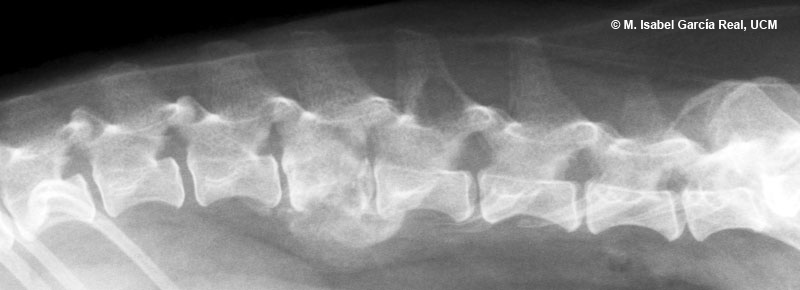
The following radiological findings are observed in the lumbar region:
- Small bony overgrowth with a regular morphology on the caudoventral aspect of the body of L2, possibly because of spondylosis deformans.
- L3 has a mixed osteoproliferative and osteolytic lesion. The osteoproliferative changes essentially affect the ventral margin of the vertebral body, while there is a moth-eaten osteolytic pattern in the vertebral body and arch.
- Narrowing of the L3-L4 intervertebral disc space.
- Geographic osteolytic lesion located in the ventral area of the spinous process of L4.


