ATLAS OF RADIOGRAPHIC INTERPRETATION

in small animals
IMAGES OF NORMAL RADIOGRAPHIC ANATOMY
ISABEL GARCÍA REAL - Diagnostic Imaging Service, Veterinary Teaching Hospital, Complutense University of Madrid - PIMCD 256 (2011-2012) - UCM

Radiological report

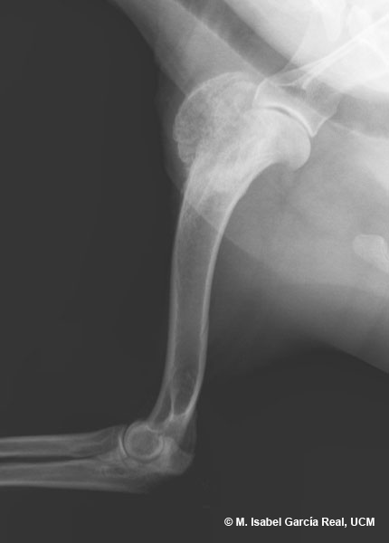
Only part of the olecranon is visible in the image, due to overexposure of the radiograph, which is digital in this case. If the radiograph is highly overexposed, the olecranon will not appear complete even if the brightness and contrast of the image are modified, and the radiograph will therefore have to be repeated with correctly adjusted exposure values. We should not mistake this artefact for a real osteolytic lesion in the ulna.

Previous

Centro Empresarial El Trovador, planta 8, oficina I - Plaza Antonio Beltrán Martínez, 1 • 50002 Zaragoza - España • Tel.: 976 461 480 • Fax: 976 423 000 • www.grupoasis.com