ATLAS OF RADIOGRAPHIC INTERPRETATION

in small animals
IMAGES OF NORMAL RADIOGRAPHIC ANATOMY
ISABEL GARCÍA REAL - Diagnostic Imaging Service, Veterinary Teaching Hospital, Complutense University of Madrid - PIMCD 256 (2011-2012) - UCM

Radiological report

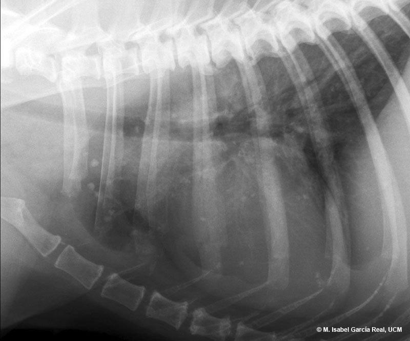
It is very unlikely that this type of image is caused by lung metastases, since they are usually not calcified, and even less in their totality (in this case, no soft-tissue opacity lesion suspicious of metastasis has been identified).


The following incidental findings are observed:

  • Spondylosis deformans affecting T4, T5, T6, T7, T8 and T9.
  • Irregular bony overgrowths at the ends of some sternebrae.
  • Calcification of bronchial walls.
Previous

Centro Empresarial El Trovador, planta 8, oficina I - Plaza Antonio Beltrán Martínez, 1 • 50002 Zaragoza - España • Tel.: 976 461 480 • Fax: 976 423 000 • www.grupoasis.com