ATLAS OF RADIOGRAPHIC INTERPRETATION

in small animals
IMAGES OF NORMAL RADIOGRAPHIC ANATOMY
ISABEL GARCÍA REAL - Diagnostic Imaging Service, Veterinary Teaching Hospital, Complutense University of Madrid - PIMCD 256 (2011-2012) - UCM

Radiological report

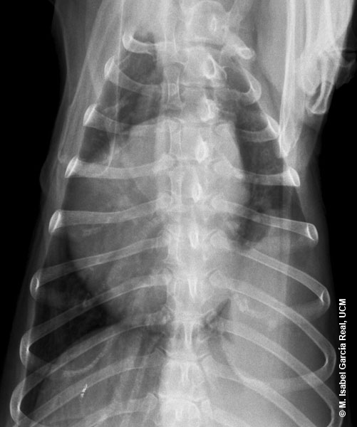
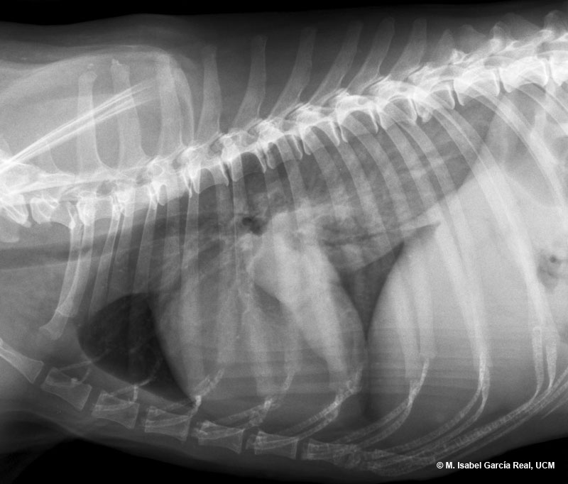
The subsequent ultrasound examination confirmed the solid ultrasound image texture of the mass and allowed the collection of a sample for cytology, which confirmed its pulmonary origin.


Computed tomography would also be indicated in this case to confirm the exact location and extension of the lesion, as well as to assess the presence of small-sized pulmonary metastases not detected on the radiographs, before planning a surgical treatment.

Previous

Centro Empresarial El Trovador, planta 8, oficina I - Plaza Antonio Beltrán Martínez, 1 • 50002 Zaragoza - España • Tel.: 976 461 480 • Fax: 976 423 000 • www.grupoasis.com