Radiological report
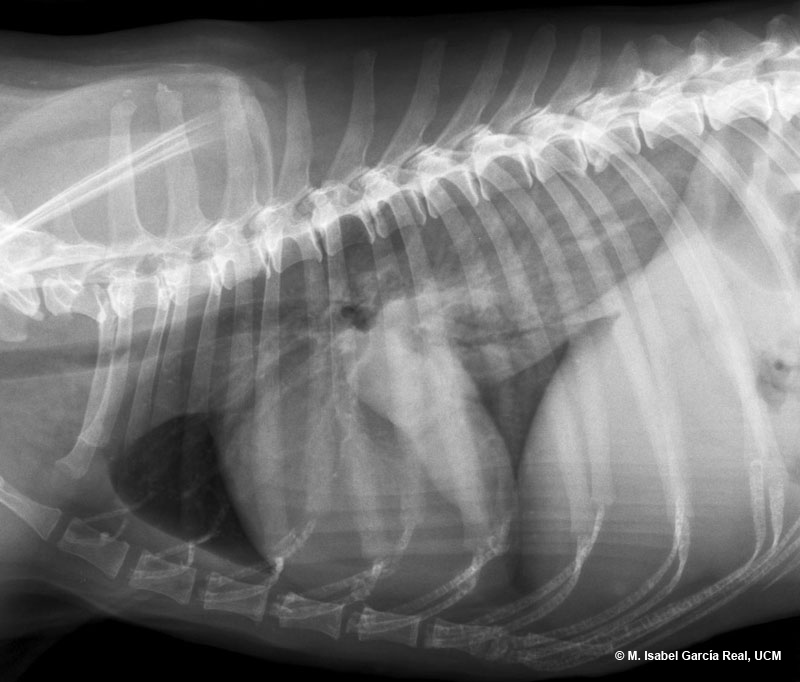
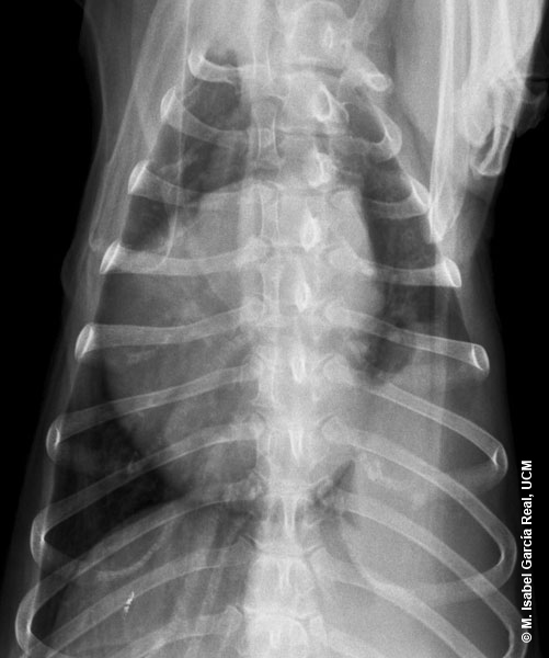
Radiographs of the thorax in right lateral and dorsoventral projections.
A soft-tissue opacity mass with a wide area of contact with the left thoracic wall (4 intercostal spaces) can be visualised in the area of the left caudal lung lobe. The cardiac silhouette appears displaced towards the right hemithorax. The margins of the mass are ill-defined on the lateral view (which could be mistaken for an area of diffuse interstitial infiltration if this was the only image available), but its medial margin is well defined on the dorsoventral view. Given the morphology and location of the mass, the pleura or lung should be considered as the most probable origins.


