ATLAS OF RADIOGRAPHIC INTERPRETATION

in small animals
IMAGES OF NORMAL RADIOGRAPHIC ANATOMY
ISABEL GARCÍA REAL - Diagnostic Imaging Service, Veterinary Teaching Hospital, Complutense University of Madrid - PIMCD 256 (2011-2012) - UCM

Radiological report

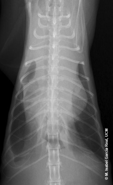
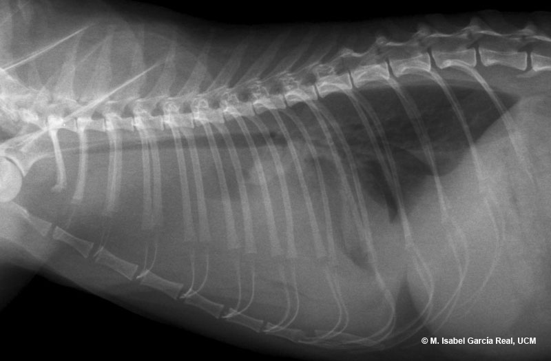
Radiographs of the thorax in right lateral and dorsoventral projections.


A marked enlargement of the shadow of the mediastinum is observed on the dorsoventral view, together with moderate distension of the medial and caudal area of the lungs, and slight retraction of the lateral margin of the right caudal lung lobe and of the dorsal margin of both caudal lobes. Most of the cardiac silhouette is masked on both views and the trachea appears displaced in dorsal direction on the lateral view.


Centro Empresarial El Trovador, planta 8, oficina I - Plaza Antonio Beltrán Martínez, 1 • 50002 Zaragoza - España • Tel.: 976 461 480 • Fax: 976 423 000 • www.grupoasis.com