Radiological report
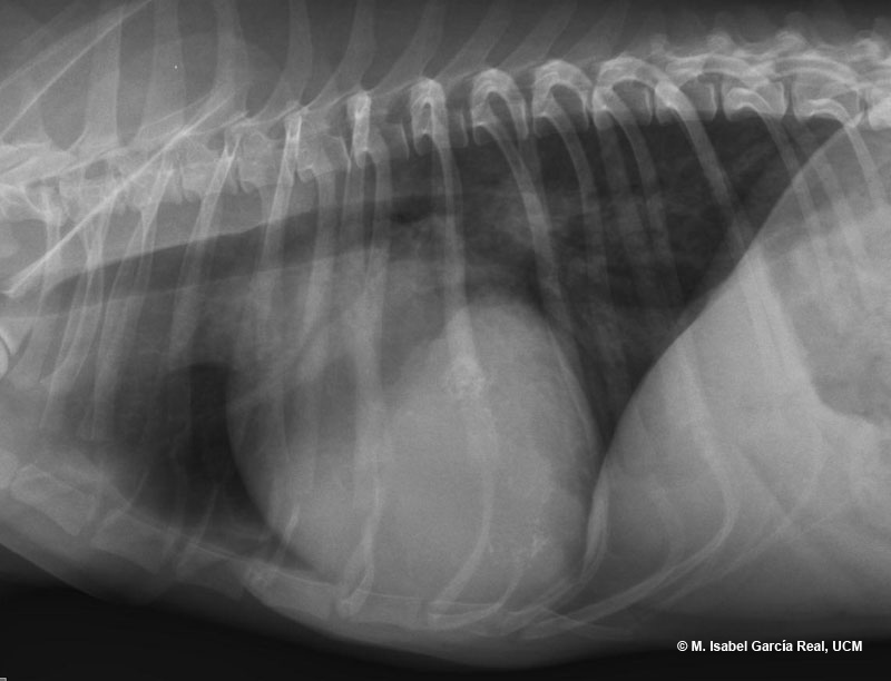
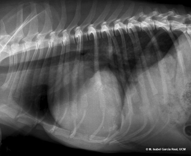
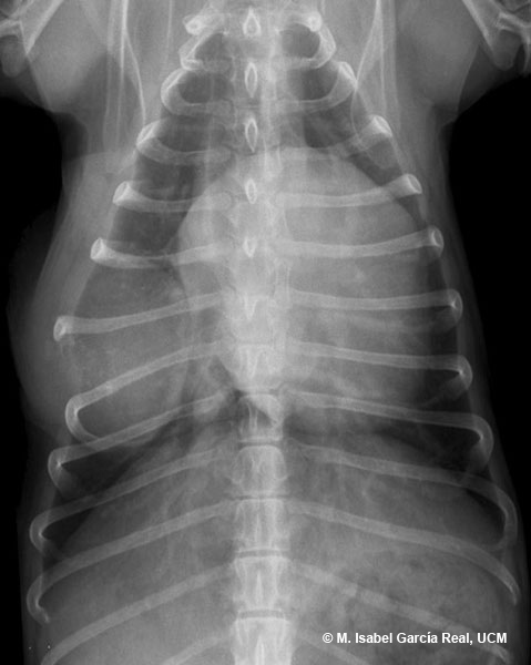
Radiographs of the thorax in right lateral, left lateral and dorsoventral projections.
Marked osteolysis is observed in the middle and distal third of the seventh right rib, which appears surrounded by a soft-tissue opacity mass with an approximately round morphology occupying the right 6th, 7th and 8th intercostal spaces. The mass has grown to a greater extent in the thoracic cavity than on the external surface of the thoracic wall. The right 8th rib appears displaced in medial and caudal direction. These findings are compatible with primary bone neoplasia of the rib, soft-tissue neoplasia secondarily affecting the adjacent rib or osteomyelitis. The last two possible diagnoses are unlikely.


