ATLAS OF RADIOGRAPHIC INTERPRETATION

in small animals
IMAGES OF NORMAL RADIOGRAPHIC ANATOMY
ISABEL GARCÍA REAL - Diagnostic Imaging Service, Veterinary Teaching Hospital, Complutense University of Madrid - PIMCD 256 (2011-2012) - UCM

Radiological report

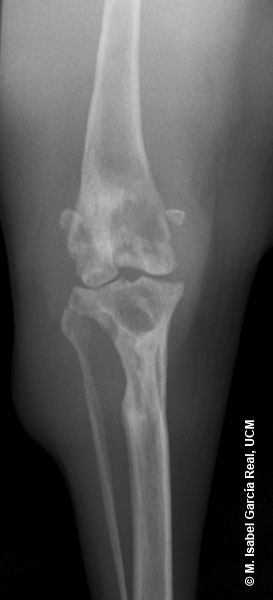
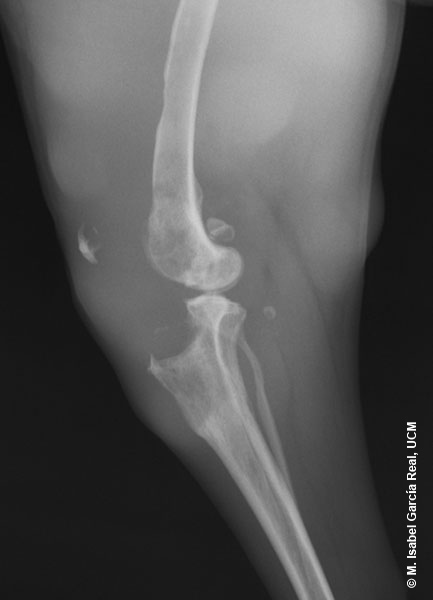
These findings are compatible with soft-tissue neoplasia secondarily affecting the adjacent bones, osteomyelitis, joint tumour (less likely as the joint surface of the femur is less affected than that of the tibia) or bone metastases.


An ultrasound examination may allow a better definition of the lesion of the soft tissues, although this assessment would be more precise using magnetic resonance imaging of the limb.

Previous

Centro Empresarial El Trovador, planta 8, oficina I - Plaza Antonio Beltrán Martínez, 1 • 50002 Zaragoza - España • Tel.: 976 461 480 • Fax: 976 423 000 • www.grupoasis.com