Radiological report
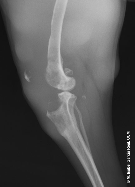
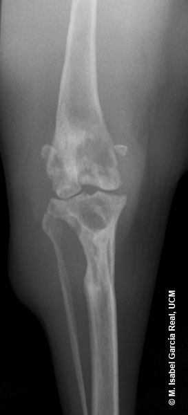
Radiographs of the stifle in mediolateral and craniocaudal projections.
Multiple osteolytic lesions with a geographical pattern are observed in the distal metaphysis of the femur, metaphysis and proximal epiphysis of the tibia and in the patella, which is displaced in cranial direction. The sesamoid bone of the popliteus muscle is also displaced, in this case in caudal direction. Irregular thinning of the cranial cortex of the femoral diaphysis and marked tumescence of the soft tissues that surround the femur and joint are also observed.


