ATLAS OF RADIOGRAPHIC INTERPRETATION

in small animals
IMAGES OF NORMAL RADIOGRAPHIC ANATOMY
ISABEL GARCÍA REAL - Diagnostic Imaging Service, Veterinary Teaching Hospital, Complutense University of Madrid - PIMCD 256 (2011-2012) - UCM

Radiological report

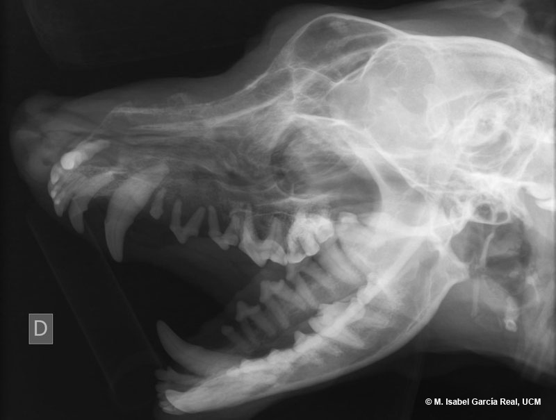
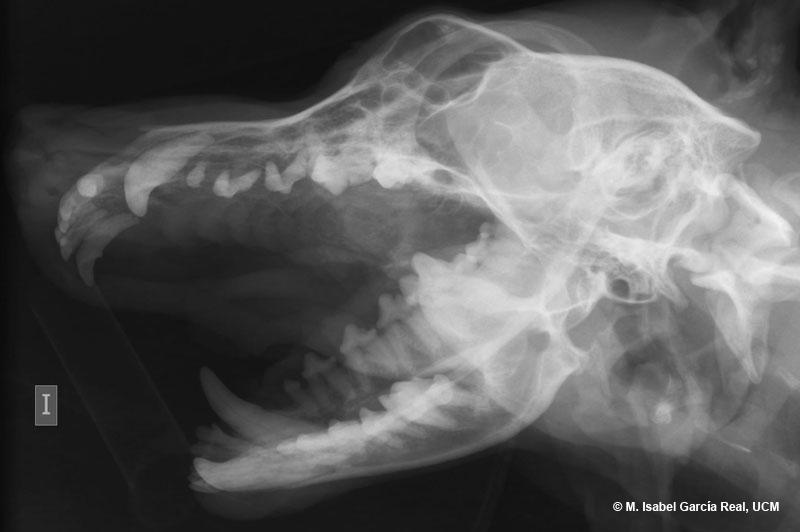
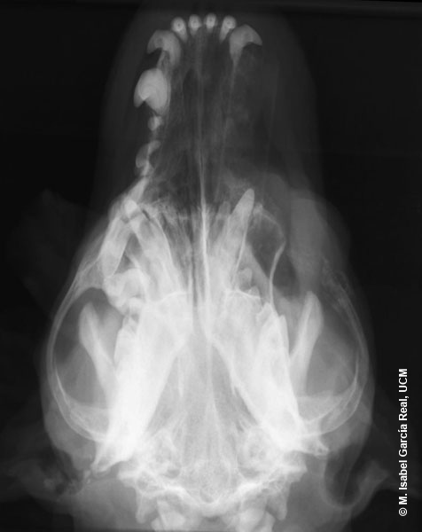
The absence of teeth can be observed on the oblique left lateral view, but the radiograph is not very useful to assess the characteristics of the lesion of the maxilla. This image is compatible with a bone neoplasm, soft-tissue neoplasm with secondary bone involvement or osteomyelitis (unlikely given the morphology and aggressiveness of the lesion).

Previous
Centro Empresarial El Trovador, planta 8, oficina I - Plaza Antonio Beltrán Martínez, 1 • 50002 Zaragoza - España • Tel.: 976 461 480 • Fax: 976 423 000 • www.grupoasis.com