Radiological report
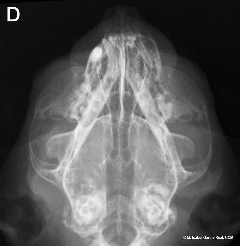
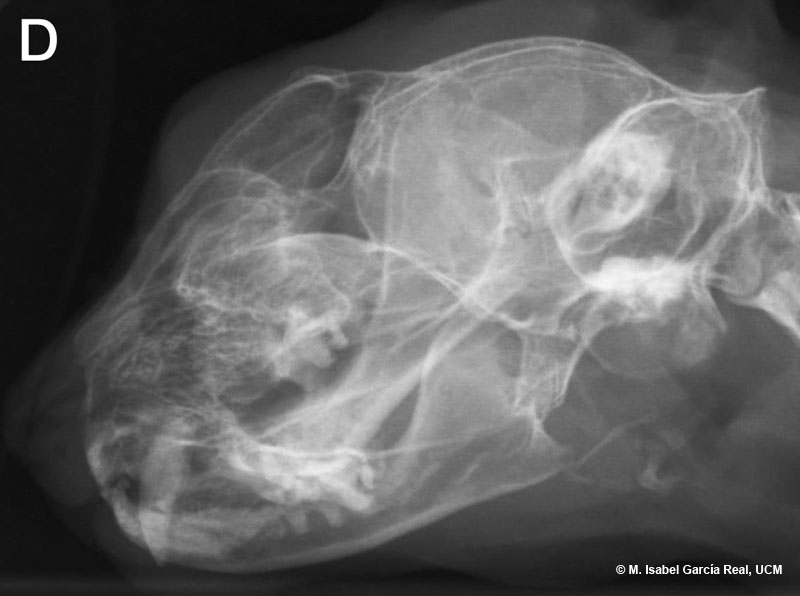
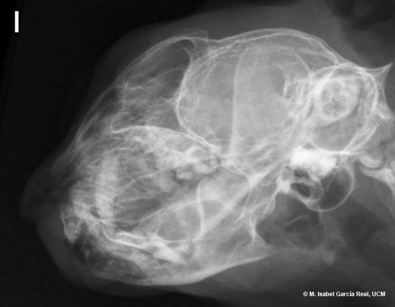
Radiographs of the head in dorsoventral, oblique right lateral and oblique left lateral projections.
Areas of osteolysis are observed in the wall of the right tympanic bulla, which also appears irregularly enlarged. A loss of the normal air content of the tympanic bulla is observed. The right ear canal appears partially obliterated. These findings are compatible with severe unilateral otitis media or soft-tissue neoplasm with secondary involvement of the adjacent bones.
As an incidental finding, the absence of numerous upper and lower teeth can be observed.


