Radiological report
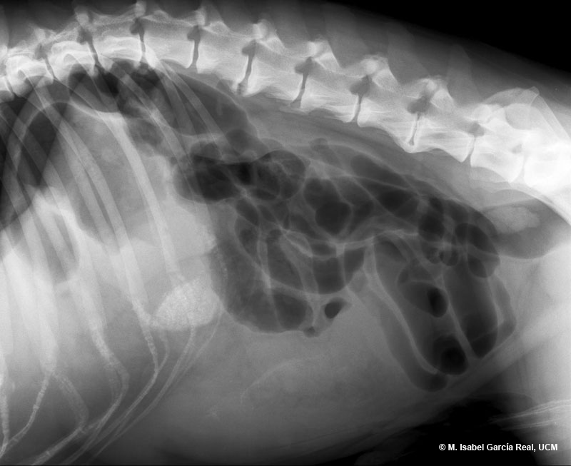
The presence of mineral density gravel in the area corresponding to the pyloric antrum (which is dorsally displaced in this case) and spondylosis deformans in T12, T13 and L1-L6 are observed as incidental findings. The L5-L6 intervertebral disc space appears reduced in width. However, given the absence of associated clinical signs, it is probably an old lesion caused by degenerative disc disease.
A marked enlargement of the spleen, which has a hypoechoic parenchyma, was observed during the subsequent ultrasound examination, as well as an absence of flow in the area of the splenic hilum and a focal peritoneal reaction around it. The presumptive diagnosis was splenic torsion, which was later confirmed during surgery.
Previous


