Radiological report
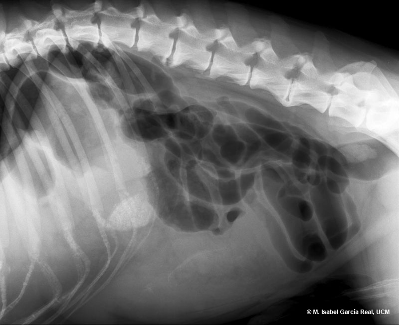
Radiograph of the abdomen in right lateral projection.
An area of soft-tissue opacity with undefined margins causing a dorsal displacement of the stomach and small intestine (mass effect) is observed in the ventral area of the mid-abdomen. Given its location, the most probable cause of this finding is splenomegaly, splenic mass or mass of another origin, in addition to ascites, peritonitis or peritoneal carcinomatosis as a cause of the loss of definition of the outline of the mass.
A generalised pattern of mild dilatation is also observed in the small intestine, and is most likely caused by a functional ileus secondary to the lesions previously described.


