ATLAS OF RADIOGRAPHIC INTERPRETATION

in small animals
IMAGES OF NORMAL RADIOGRAPHIC ANATOMY
ISABEL GARCÍA REAL - Diagnostic Imaging Service, Veterinary Teaching Hospital, Complutense University of Madrid - PIMCD 256 (2011-2012) - UCM

Radiological report


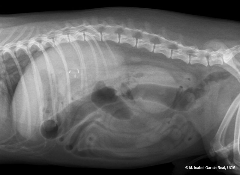
The mass is subsequently assessed by ultrasound examination. It is located in the peritoneum and is not attached to adjacent organs. An echoic wall and anechoic content with suspended echoes (compatible with fluid rich in cells) is observed in the mass, which suggests peritoneal abscess as the most probable presumptive diagnosis. This was confirmed with the subsequent surgery.

Previous

Centro Empresarial El Trovador, planta 8, oficina I - Plaza Antonio Beltrán Martínez, 1 • 50002 Zaragoza - España • Tel.: 976 461 480 • Fax: 976 423 000 • www.grupoasis.com