ATLAS OF RADIOGRAPHIC INTERPRETATION

in small animals
IMAGES OF NORMAL RADIOGRAPHIC ANATOMY
ISABEL GARCÍA REAL - Diagnostic Imaging Service, Veterinary Teaching Hospital, Complutense University of Madrid - PIMCD 256 (2011-2012) - UCM

Radiological report


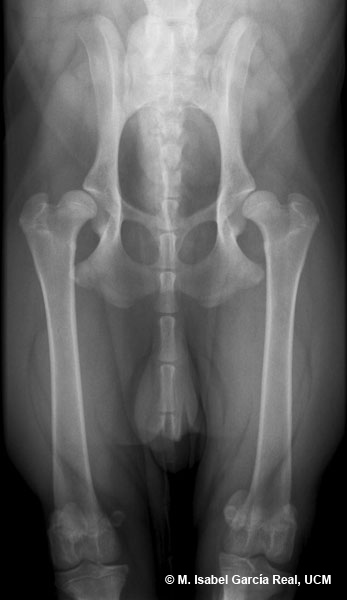
Radiograph of the hip in ventrodorsal projection.

Marked subluxation of both femoral heads is observed, with a percentage of acetabular coverage of 20 %. The Norberg angle has a value of 77º in the right coxofemoral joint and 76º in the left joint. Both acetabula appear moderately flattened.


Centro Empresarial El Trovador, planta 8, oficina I - Plaza Antonio Beltrán Martínez, 1 • 50002 Zaragoza - España • Tel.: 976 461 480 • Fax: 976 423 000 • www.grupoasis.com