ATLAS OF RADIOGRAPHIC INTERPRETATION

in small animals
IMAGES OF NORMAL RADIOGRAPHIC ANATOMY
ISABEL GARCÍA REAL - Diagnostic Imaging Service, Veterinary Teaching Hospital, Complutense University of Madrid - PIMCD 256 (2011-2012) - UCM

Radiological report

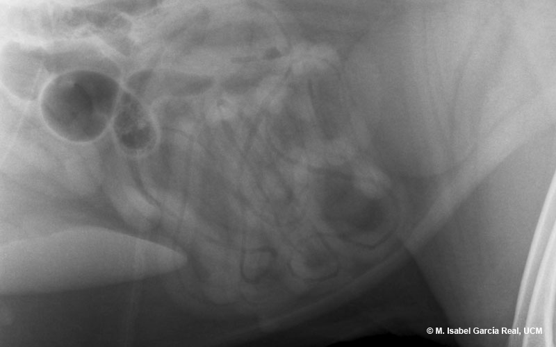
An enlarged image of the central area of the abdomen in lateral projection is shown.


The image pattern of the small intestinal loops is compatible with a small amount of gas content and a larger amount of fluid/mucous content, which creates a false visual effect of increased thickness of the walls. It should be remembered that the thickness of the walls of the stomach and intestine can only be assessed by means of ultrasonography.

Previous

Centro Empresarial El Trovador, planta 8, oficina I - Plaza Antonio Beltrán Martínez, 1 • 50002 Zaragoza - España • Tel.: 976 461 480 • Fax: 976 423 000 • www.grupoasis.com