Radiological report
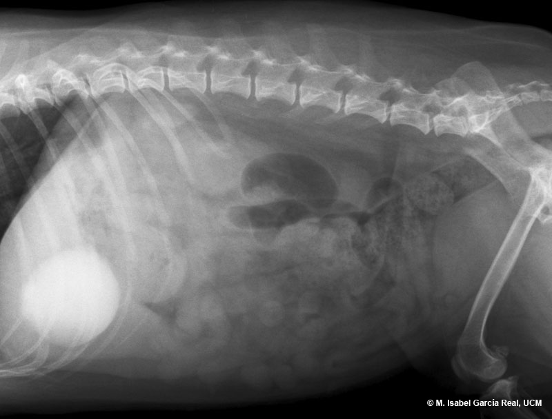
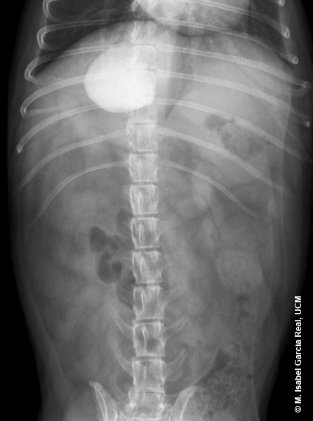
Radiographs of the abdomen in right lateral and ventrodorsal projections.
A mineral density shadow is observed in the cranial area of the abdomen, with an approximately round morphology, a poorly defined medial margin and a diameter of 2 intercostal spaces. There is no displacement of adjacent structures. This image is compatible with sludge or a large-sized calculus in the gallbladder. Although a gastric foreign body may be suspected due to the close location of the shadow to the stomach, it is clear that it is located adjacent to it on the lateral view.
The presence of a large amount of gallbladder sludge was later confirmed by means of ultrasonography.
Previous


